
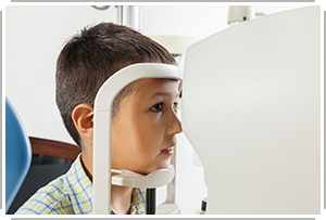
Neurologic Assessment
 Courtesy ThinkStock: Dangubic
|
It is also important to do a thorough neurologic exam. Most of the time, neurologic testing is used to rule out worse issues, rather than to rule in concussion.
Neurologic assessment typically involves a head-to-toe exam with components such as:
|
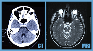
To Scan or Not to ScanNeuroimaging will not provide a solid "This is a concussion" diagnosis, but is useful in ruling out structural abnormalities when indicated. Along with concerns about structural abnormalities in an acute exam, other red flags for neuroimaging include focal neurologic deficits and/or an atypical course - for example, a sudden increase in symptoms for no reason. |
 Courtesy ThinkStock: nimon_t & popovaphoto
|
This statement from the American Society for Sports Medicine can equip you with facts to counter any arguments from concerned family members or other lay people who may question the decision to forego neuroimaging.
From the 2013 American Medical Society for Sports Medicine Position Statement:
Concussion in Sport
"The vast majority of athletes with sports-related concussion do not require neuroimaging. Standard neuroimaging with computed tomography (CT) or magnetic resonance imaging (MRI) is negative in concussion, but is used to evaluate for more serious brain injury."
 |
 |
Best used acutely for evaluating for:
|
More sensitive for:
|